BIOPSYCHOLOGY I (BPS108)

Find out how our minds and brains relate to one another

Stress, anger, concentration, clarity of thought, etc. are all influenced by your physical wellbeing (eg. fitness, general health, the food you eat). This course develops an understanding of the relationship between our brain and nervous system with our thoughts and behaviours. It examines how a person's physical body is associated with their mental processing.

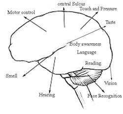
Learn about the biology of the brain and how it relates to things like our senses, the way we perceive the world, and altered states of consciousness.

Graduate Comment: "I thoroughly enjoyed the course and found ACS to be wonderful in all aspects" D. Kenyon, ACS Biopsychology student

Learn about the Mind-Body Relationship

Biopsychology brings together biology and psychology. It is the study of where the physical structures in the brain are located and how they are involved in the way we experience the world around us. 

In this course, students learn about how messages are transmitted inside the brain and body, the role of chemicals and hormones, and the detection and interpretation of sensory information.

The course also examines the roles of hormones and chemical messengers in bodily systems, ethical considerations in research, sexual behaviour, the impact of stress, the psychology of emotions, and the brain and consciousness.

Who needs to understand biopsychology?

Anyone who deals with people will enhance their understanding of the people they deal with through an understanding of biopsychology.

People involved in welfare services, counselling and child services (e.g. teachers, youth leaders, etc.) in particular can find a greater understanding of this subject to be very useful). Develop your ability to comprehend the way in which a person's state of mind influences their physical body; and the way in which their physical body affects their state of mind.


Course Structure and Contents

There are eight lessons in this module as follows:

  1. Introduction
    Types of external and internal stimuli, mind-body debate, introduction to the nervous system.
  2. The senses
    Sensory input, sensory perception, description of the major senses.
  3. The Nervous System
    Description of the neurons, the central nervous system, peripheral nervous system, including the autonomic nervous system.
  4. The Endocrine System
    Effect of hormones on behaviour and physiology, association of endocrine system and nervous system, connection between external and internal stimuli.
  5. Stress
    Types of stressors, physical affects of stress, personality & stress.
  6. Emotions
    Homeostasis, eating disorders, physiological responses to emotions, theories of emotion.
  7. Consciousness
    Degrees of consciousness, awareness & attention, altered states of consciousness.
  8. Project


Duration:
100 hours


RESEARCH METHODS OF BIOPSYCHOLOGY


Neuroanatomical Techniques

There are many different techniques used in research in biopsychology. These are only a few of them. Many of these techniques make use of studies on animals. The ethics of animal research is discussed further in the ethics section of this lesson.

Histological Procedures - Tissue preparing techniques. We cannot see the cellular detail of the brain by just looking at it, and even with a microscope we need fixation and staining of the neural tissue.

Fixation - To study an organism as it was at the time of death, we must destroy the autolytic enzymes (self-dissolving enzymes), which cause tissue to decompose. The tissue is preserved by use of a fixative, such as formalin (the aqueous version of formaldehyde - a gas)

Staining - Without staining, no details of the brain are revealed. The study of neuroanatomy requires special histological stains. The basic types used are - cell-body stains, myelin stains, membrane stains and degenerating-axon stains.

Tracing Neural Pathways - Staining does not help to show the detail of neurons, as a tangle of neurons is shown. Special techniques are required to make the connections being studied stand out from all others.

Degenerative Studies - When a cell body is destroyed, the distal portion of the axon quickly dies and disintegrates - this is called anterograde degeneration. A degenerating-axon stain will identify the dying axons as trails of black droplets.

Amino acid autoradiography - Amino acids are transported via special means cause axoplasmic flow. This technique uses the axoplasmic flow. A researcher will inject radioactive amino acids into the brain of an animal and allows the animal to live for a couple of days. The cell bodies will take up the radioactive amino acids and incorporate them into proteins. The animal is then killed and the brain studied. The radioactive proteins show up as black spots.

Horseradish Peroxidase (HRP) is an enzyme - a protein capable of splitting certain peroxide molecules and turning them into insoluble salts. HRP is injected into the brain. It eventually reaches the cell bodies of neurons that send axons into the region of the brain that received the injection. After a day or so, the animal is killed and the brain studied. The HRP technique permits identification of neurons that project axons to a particular area.

These techniques can show us the source of inputs to particular parts of the brain and the location to which axons are sent. So the interconnections of some parts of the brain can be studied with great accuracy.

Studies with Living Brains

BRAIN SCANS can study cortical functioning. They include:

CAT scans (computerised axial tomography): An x-ray beam goes through an individual's head and a level of radioactivity is detected. The level is lower when the X-ray passes through dense material. CAT scans are useful for detecting tumours, brain abnormalities and clots. They do not show precise locations of brain damage or show the actual functioning of the brain. They are also very expensive.

CAT scans (below) provide 3D images for easy observation of brain abnormalities.

PET scans (positron emission tomography): Shows the brain in action and also what part of the brain is active in certain tasks. It only shows activity over a 60 second period. PET scans display differences in brain regions through blood flow and fuel metabolism. Differences can be observed between different individuals and within an individual both in terms of time and brain region. A radioactive tracing substance is administered either into the blood stream via injections or inhalation, or by administering an artificial substance similar to glucose (a brain fuel), then the activity in different brain regions is observed. Areas involved in motor control and sensory stimulation can be observed.

MRI scans (magnetic resonance imaging): Produce clearer and more detailed pictures than CAT scans. MRI is used to detect structural details of the brain. Some substances contained in water molecules in the brain, such as hydrogen atoms, respond to magnetic fields. When a magnetic field is applied, the interaction between molecules and this field are monitored, allowing different tissues to be distinguished, based on their constituent molecules. These changes are interpreted by a computer. They can be used to detect very small tumours. They can still only tell us about the structure of the brain rather than its functioning.

Functional MRI: This produces images of the brain with areas of high activity indicated, so we can get a picture of the brain whilst functioning. This provides more spatial information than PET scans and shows changes over shorter periods of time.

Squid magnetometry (superconducting quantum interference devices): This measure the magnetic field produced by neuron activity in the brain. Irrelevant magnetism may interfere with results and the machine has to be kept at extremely low temperatures.


How This Course Could Help You

This course may be studied by itself or as part of a certificate or higher level course. The course will be of most use to those with an interest in:

  • Psychology
  • Psychotherapy
  • Teaching
  • Research
  • Biological sciences
  • Health sciences
  • Health professions


Get started today and make a difference!

Simply click on the ENROL NOW icon at the top of this page